
COAT COLOR
This page was created for a reference for breeders interested in gene theory, as it pertains to the Blue Lacy breed. This information is a highlight of essential components involved in coat color for the Blue Lacy. Readers are encouraged to research further books and articles on Canine Genetics, if they desire to know more about this subject.This being said, canine coat color is still a field of study that is only partially understood. None of the studies or published accounts has resulted in a complete genetic system, capable of fully determining canine coat colors. This article is designed to provide information in layman’s terms, for easier reading.
Coat Color Appearance
The Blue Lacys’ coat is predominately solid, or solid with points in appearance. White markings may also appear, most commonly on the toes and chest (brisket) regions.
Color Categories
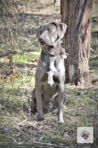

Blue – This color designation ranges in shades of light grayish or light chocolate in appearance, to dark gunmetal gray or charcoal.
Red – The color range for Reds, includes all shades between cream and dark red. They can present minimal slight blue coloration that shows through.
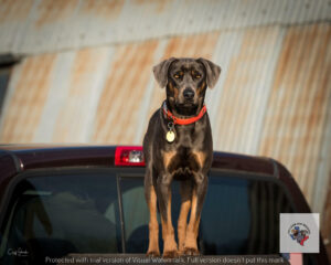
Tri – Tri’s are Blue with Red markings on the points (Red points: over the eyes, on cheeks, under the tail around the anal area, and down the legs.) The Blue and Red markings are subject to the variation of shades of color that apply within those categories. Note: If a Tri looses, or is not born with the head dominantly blue, which gives a saddle back appearance, this needs to be documented, and reviewed before becoming part of the standard.
White Markings
White on the chest region can extend up to the chin and down to the underside of the torso. White on the toes can extend to what would be referred to as a white sock, but preferably below the “ankle” joint. Dogs may also exhibit minimal white hairs at the tip of the tail. White on the muzzle above the chin, the head, or the front legs higher than one inch above the “ankle” joint and/or the top of the neck region is disqualified for breeding purposes.
Note: No documented Blue Lacy has had excessive white on its back legs, reaching above the hocks. The breed has, however, occasionally displayed any of these other white patterns.
The science behind “Predominantly Solid or Solid with Points Color Appearance” and “White Markings”
At the embryo stage of canine development, special cells are involved in the formation of the central nervous system (the brain and spinal cord). These special cells, left over at the edge of the nervous system formation, become Neural Crest Cells. Neural crest cells migrate throughout the body to form the skin melanocytes and much more. The gene that produces white patches on a dog is believed to interfere with the number of neural crest cells produced. It may also interfere with the neural crest cell’s ability to migrate. Yes, all this starts at embryo formation!
Where melanocytes are present, color can be produced. These melanocytes serve as a shield to protect deeper structures such as organs from harmful ultraviolet rays. Melanin is produced by melanocytes, and is responsible for hair and skin color. White markings are the result of a lack of melanin “pigment” in an area.
In breeds with primarily solid colors or solid with points, such as the Blue Lacy, their pigment distribution is even. When pigment is absent in an area, white patches appear.
Distribution of Color in Coat
In this section I would like to refer to a helpful web page that already exists on the topic. The link provided directs you to the page that refers to the coat colors associated with the Blue Lacy Breed. http://homepage.usask.ca/~schmutz/dilutions.html
Recognized Dog Color Series
A (agouti), B (brown), C (albino/chinchilla dilute), D (dilution), E (extension), G (graying), M (merle), R (roaning), S (white spotting) and T (ticking).
Color gene series recognized in the Blue Lacy breed could consist of:
- “A” (Agouti) – Primary Coat Color – The Agouti series has three alleles in this group. This series has multiple alleles, which influence the amounts of dark and light pigment in the complete coat hairs.
- “B” (brown) – This color series has two alleles. Possible colors within this series, red dilute, and black lightened to chocolate. Nose leather color will be brown to chocolate in this series.
- Note: This is not prominent in the Blue Lacy, if seen at all.
- “C” (chinchilla dilute) – This series has several alleles. Many believe there are more than the defined alleles commonly listed for this gene. Suggestions of minor variants on the c-ch allele – “C” allows full pigmentation such deep reds, and blues. “C” suppresses black and red pigments to silvery gray and cream (off white).
- Note: This series could be the reason for the variations in “shades” seen within the Blue Lacy breed, but that is undocumented at this time. This series does not affect the color of the nose leather.
- “D” (dilution) – The Dilution Series has two alleles. This series acts with other loci to cause different expressions of color genes, but does not actually cause color.
- Note: This is believed to be the most common gene-effecting coloring seen in the Blue Lacy breed.
- “E” (extension) – This series has two alleles.
- Note: If there is a contributor to color for certain “Red” colored Blue Lacys, it would be the recessive “E”. Extensions are gold, yellow and cream.
- “T” (ticking) – This series also has two alleles. – Colorflecks seen in white areas. This series is not always seen at birth, but can appear as a dog ages. The color that shows through the white will be the base color. It is common for ticking to result in only changing the color of the skin and not affecting the color of the white hair in many breeds.
- Note: This is possible with the Blue Lacy breed as well, but would be more of a skin color effect on older dogs.
- “S” (spotting) – The White Spotted series has four alleles.
- Note: This gene is expressed in the Blue Lacy breed and the next section will expand on this.
- “S” White Spotted Gene
- Understanding the white spotted gene’s presence within the Blue Lacy breed is of importance. As mentioned above, melanocytes migrate down from the spinal column during embryo formation. In dogs, it is not uncommon for actual “S” solid color dogs, otherwise Blue or Red in color, to have white toes, chest, or tip of the tail. Some believe this can be the result of random events, other than a specific allele. It will take place very late in the embryo’s development to prevent the melanocytes from being distributed to these areas completely as a result of the random event. There are also beliefs, and speculations, that the rate of melanocyte distribution and migration itself may be inherited.
- Studies indicate small amounts of white on the chest, toes, or tip of the tail; they do not seem to be the cause of MITF mutations. You ask, what is MITF? MITF stands for Microphthalmia Associated Transcription Factor. It was one of the first genes identified, published in 2007, which causes at least some spotting patterns and/or potential mutations causing some forms of white spotting in coat color.
- The major series of genes that distribute pigment in dogs’ coats is called the “S” or spotting series. This series controls the amount of white markings expressed.
- S – Solid color. Modifying factors – white on toes, chest and tail tip.
- Note: This applies to the Blue Lacy Breed
- si – Irish spotting – Definite Pattern – white on muzzle, chest, neck, legs and tail. Modifying factors control the amount of white seen. “Plus” factors extend the white and area, while “minus” factors reduce the white.
- Note: This applies to the Blue Lacy Breed.
- sp – Piebald – 50 % white covers most of body
- Note: Not a factor in the Blue Lacy breed.
- sw – Extreme Piebald – Produces an all white dog
- Note: Not a factor in the Blue Lacy breed.
Charles Little (1957) suggested that most spotting was caused by the four “S”alleles listed above. This does not fit with recently published DNA studies. Ojvind Winge (1950) does not assign alleles to the Spotting Series in his book. Mr. Winge states that there is a recessive allele that controls “white-mottling”, and when it is homozygous, the dog will exhibit more white. When the recessive allele isn’t present, he suggests white markings are minimal, or not expressed. MINIMAL, is implied to be represented on the feet, tail tip and chest.
How should breeders look at breeding concerning excessive white guidelines?
During conception, reproductive cells of the parents unite into a replication type, through which individual breed characteristics are transmitted. Puppies receive half of their chromosomes from the dam and the other half from their sire.
Blue Lacy owners may have a certain look or white pattern they desire. Accountable breeders will only breed parents that conform to breed standards, and exhibit an excellent temperament, health, type, character, working ability and drive. A particular color, or the amount of white exhibited, within breeding standards, should be considered after all other traits.
Now, for the white spotted gene: Again, after all the main breeding traits are compared, if a breeder wants to reduce the risk of pups being born with excessive white, they should select a sire and dam that complement each other. If a breeder feels that a sire or dam has a large amount of white present, but still within breeding standards, they should select an opposite sex Blue Lacy to breed this dog with, which has less white showing. However, this is not a foolproof method, as there are many factors that play a role in pigment distribution.
There are documented litters where both parents have minimal white markings, and they still produce pups with excessive white coloration. There is also documentation of litters in which both parents have large white areas, although still within breeding standards. All the pups exhibit less white than the parents, and in a different distribution on the body.
Dog breeds, coat colors, and markings are only described by what we see in front of us. We look at the dog’s phenotype, or appearance. We cannot readily visualize the genetic makeup, called the genotype. Knowing there can be a difference in a dog’s phenotype versus his genotype, we can only do the best we can as breeders. This means figuring percentage and probabilities before a mating takes place.
Just because a litter mate, or mates, has excessive white for the breed, that does not affect other pups in the same litter. These pups do not exhibit excessive white. Those pups were born with, and possess, the gene that expresses pigment distribution to meet the breeding standard. These individual pups have a high enough percentage rate of standardization to produce breeding standard pups, when bred to another dog meeting the standard.
Pups that would be classified as having excessive white are still just as pure blood Blue Lacy as their littermates. There may be nothing wrong with their drive or abilities to hunt, and/or be a companion dog. There are several benefits to having these pups in Blue Lacy breeding. Many individuals looking to obtain a Blue Lacy are not interested in breeding. Individuals that plan on spaying and neutering make exemplary owners for pups with excessive white markings. These pups with extra white, or pups that may not meet the standard for other reasons, can fill that slot. The new owners can enjoy and love them as much as they would a breeding standard pup from the same litter. This leaves more breeding standard pups in the breeding program.
The white spotted gene will always be a part of the Blue Lacy. Trying to breed it out would not maintain the breed, or the standards by which it has always been identified.
The Greyhound also possesses the white spotted gene, but excessive white was not an issue for breed standards within Greyhounds. You can look at a Greyhound, no matter what color it is, and identify its breed by their body structure.
The only reason for having excessive white as a disqualification for the breeding standard Blue Lacys, is to set it apart from other dog breeds. These breeds share a similar body type, and allow more white in their standard. Color and markings are maintained for breed identification only.
There is much more information, and studies on Coat Color and genetics. This article expresses the author’s opinion, based upon observations and a thirteen-year study of coat color and genetics within the Blue Lacy breed. It was written to help breeders become more aware of color genetics within this breed. Several studies and books have been published on this topic.
Below is a list of recommended research materials.
Animal Genetic:
- Internet articles by Sue Ann Bowling, http://bowlingsite.mcf.com/GENETICS/Genetics.html
- Dog Coat Color Genetics, Sheila M. Schmutz, PH.D., Professor, http://homepage.usask.ca/~schmutz/dogcolors.html
- Dog Coat Color Genetics, J. Chapell’s , http://abnormality.purpleflowers.net/genetics/index.htm
- Canine Coat Color – Inheritance and Appearance (coat colors and coat color inheritance in dogs) with an emphasis on colors in Borzoi, 1995 Bonnie Dalzell, MA, Version 8-21-97, http://www.borzois.com/coat.color/coat.color.html
Studies related to Diluted Coat Color:
Philipp U, Hamann H, Mecklenburg L, Nishino S, Mignot E, Schmutz SM, Leeb T. 2005. Polymorphisms within the canine MLPH gene are associated with dilute coat color in dogs . BMC Genetics 6:34-. (This article is published in a publicly accessible online journal at http://www.biomedcentral.com/1471-2156/6/34)
Studies related to the “White Spotted” gene:
- Brenig, B., Pfeiffer, I., Jaggy ,A., Kathmann, I., Balzari, M., Gaillard, C. and Dolf, G. 2003. Analysis of the 5? region of the canine PAX3 gene and exclusion as candidate for Dalmation deafness. Animal Genetics 34: 47-50
- Krempler, A., Breen, M. and Brenig, B. 2000. Assignment of the canine paired-box 3 (PAX3) gene to chromosome 37q16->q17 by in situ hybridization. Cytogenet. Cell Genet. 90 (1-2), 66-67.
- Metallinos, D and Rine, J. 2000. Exclusion of EDNRB and KIT as the basis for white spotting in Border Collies. Genome Biology (a web based only journal) online article
- Schmutz S.M., Moker J.S, Yuzbasiyan-Gurkan V., Zemke D., Sampson J., Lingaas F., Susana Dunner S., and G Dolf. 2001. DCT and EDNRB map to DogMap Linkage Group L07. Animal Genetics 32:321.
- Schmutz, S. M., T. G. Berryere, and C. A. Sharp. 2003. KITLG mapping to CFA15 and exclusion as a candidate gene for merle. Animal Genetics 34: 75-76.
- Zemke, D. and V. Yuzbasiyan-Gurkan. 1999. A single nucleotide polymorphism and a (GA)n microsatellite in intron 6 of the canine endothelin receptor B (EDNRB) gene. Anim. Genet. 30:390.